
Peripheral Giant-cell Granuloma Associated with Peri-implant Tissues
Clinical Case Report
 Home
Home
Clinical Case Report
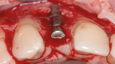
A 35-year old female patient was referred to the Department of Oral Surgery and Stomatology at the University of Bern, Switzerland, for examination of an implant site that had exhibited clinical signs of slightly delayed wound healing. In addition, the referring clinician found no evidence for a facial bone wall when she raised a flap to gain access to the implant for abutment connection. Four months earlier, she had inserted a bone-level implant in a single-tooth gap, where the lateral incisor had been extracted due to a chronic periapical lesion on the mesial aspect of the root. Implant placement was combined with simultaneous bone augmentation using deproteinized bovine bone mineral (DBBM, Bio-Oss®; Geistlich, Wolhusen, Switzerland) and a collagen membrane (Bio- Gide®; Geistlich), followed by primary wound closure. The patient also provided the postsurgical radiograph that displayed the implant with a 3.5-mm healing cap.
Recommended content
Download the QR code with a link to this page and use it in your presentations or share it on social media.
Download QR code


