Case

5.0The average rating is 5 stars out of 5.

TG14 - Immediate Implant Placement and Immediate Provisionalization with a Prefabricated-Shell Provisional Crown

Arndt Happe

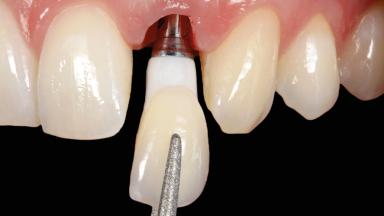
A healthy 31-year-old female patient presented with a failing maxillary left lateral incisor crown. The crown regularly loosened, and the remaining tooth was neither restorable nor rational to treat. The patient had a high smile line, a medium soft tissue biotype with a compromised mesial papilla (shorter than the contralateral one), and a horizontal scar in the buccal soft tissue as a result of past periapical surgery. Probing depths at the failing tooth and adjacent teeth were normal, in the range of 2–3 mm. The full-mouth bleeding score was 0% and the full-mouth plaque score was below 30%.

Surgical classification
Complex
Prosthodontic classification
Complex
Source
Treatment Guide 14
Purchase price
10 Academy Coins
CPD/CME
0.25 hours

General Risk Assessment

Patient-related Factors
Smoking Habit None
Oral hygiene Good
Compliance Good
Patient's Expectations Realistic
Patient-medical Factors
Medical Status Healthy, uneventful healing
Medical Fitness Healthy, able to undergo planned anesthesia and surgical procedure (ASA I)
Medications No medications that would negatively affect the surgical procedure and outcomes.
Radiation Treatment None
Growth Status Complete
Site-related Factors
Periodontal Status No history of periodontal disease, or any active periodontal disease.
Access Adequate
Pathology near the implant site None
Previous surgeries in planned implant site Previous procedures resulting in none or minimal bone and soft tissue changes.

Surgical Classification

Surgical Complexity
Timing of placement Immediate Placement (extraction sockets) (Type I)
Simultaneous or Staged grafting procedures Implant placement with simultaneous hard and soft tissue procedures
Anatomy
Bone Volume - Horizontal Adequate
Bone Volume - Vertical Adequate
Keratinized Tissue Minimal (2-4 mm)
Soft Tissue Quality Presence of minimal scars/no inflammation
Proximity to vital anatomic structures Minimal risk of involvement
Adjacent Teeth
Papilla Deficient
Recession Absent
Interproximal attachment At CEJ
Extractions
Radicular morphology Uniradicular
Available apical bone to achieve primary stability Sufficient height ( ≥ 4 mm) and width (> 2 mm around apex of planned implant)
Socket walls Intact
Thickness of buccal wall less than 2 mm
Anticipated residual defect after implant placement 2 mm or less

Prosthodontic Classification

Complicating Factors
Biological Screw-retained restorations with appropriate contours
Mechanical/Technical Absence of contributing factors
Prosthesis Factors
Prosthetic volume Adequate. Space available for ideal anatomy of the restoration
Inter-occlusal space Adequate. Capable to create an anatomically & functionally correct planned restoration
Volume and characteristics of the edentulous ridge (fixed) Adequate. No adjunctive therapy or prosthetic soft tissue replacement will be necessary
Esthetic Factors
Gingival display at full smile High
Shape of tooth crowns Triangular
Restorative status of neighboring teeth Virgin
Gingival Phenotype Medium-scalloped, medium-thick
Bone level on adjacent teeth ≤5 mm to contact point
Occlusal Factors
Occlusal scheme User-defined occlusal scheme achievable
Involvement in occlusion Minimal or no involvement
Occlusal parafunction Absent
Complexity
Loading Protocol Immediate
Implant-supported provisional restoration Required, elevated esthetic and/or functional demands
Timing of placement Immediate Placement (extraction sockets) (Type I)

Esthetic Risk Assessment

Esthetic Risk Assessment
Medical Status Healthy, uneventful healing
Smoking Habit None
Gingival display at full smile High
Width of edentulous span 1 tooth (≥ 7mm, standard diameter implant) 1 Tooth (≥ 6mm, narrow diameter implant)
Shape of tooth crowns Triangular
Restorative status of neighboring teeth Virgin
Gingival Phenotype Medium-scalloped, medium-thick
Infection at implant site None
Soft tissue anatomy Soft-tissue defects
Bone level on adjacent teeth ≤5 mm to contact point
Thickness of buccal wall less than 2 mm
Patient's Expectations Realistic
Publication date: Oct 27, 2023 Last review date: Oct 20, 2023 Next review date: Oct 20, 2026

ITI QR code Mvc

Share this page

Download the QR code with a link to this page and use it in your presentations or share it on social media.

Download QR code
QR code