Early Implant Placement with Simultaneous Contour Augmentation using GBR - Clinical Case Video - Home
 Home
Home
Clinical Case Video
Early Implant Placement with Simultaneous Contour Augmentation using GBR
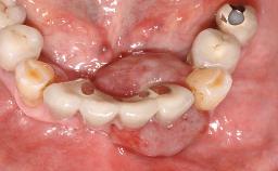
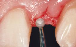
Surgical treatment of a 32-year old, healthy and non-smoking female who required removal and replacement of a lateral maxillary incisor due to internal root resorption. The inflammatory process caused a reduction of the crestal bone level on the distal side of the tooth necessitating an augmentation procedure to meet the patient's high esthetic demands. Due to the high smile line, the thin soft-tissue biotype and triangular-shaped teeth several esthetic risk factors are present.
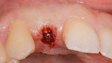
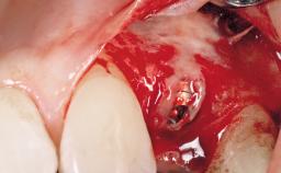
After flapless tooth removal and a healing period of 6 weeks a diameter-reduced two-piece implant is placed. The bone defect on the facial aspect is corrected with a contour augmentation using autologous bone chips covered with DBBM particles and a collagen membrane according to the Guided Bone Regeneration (GBR) approach.
Produced in cooperation with CCDE and Video-Atelier Keel.
- Surgical SAC classification
- Advanced
- Prosthodontic SAC classification
- Advanced
- Duration
- 18 minutes
- Purchase price
- 18 Academy Coins
- CPD/CME
- 0.3 hours
Recommended content
Share this page
Download the QR code with a link to this page and use it in your presentations or share it on social media.
Download QR code